Nebraska Animal Medical Center
Diagnostic Ultrasound
An ultrasound is a highly useful tool when evaluating heart conditions, internal organs, cysts and tumors.

Why Would My Pet Need an Ultrasound?
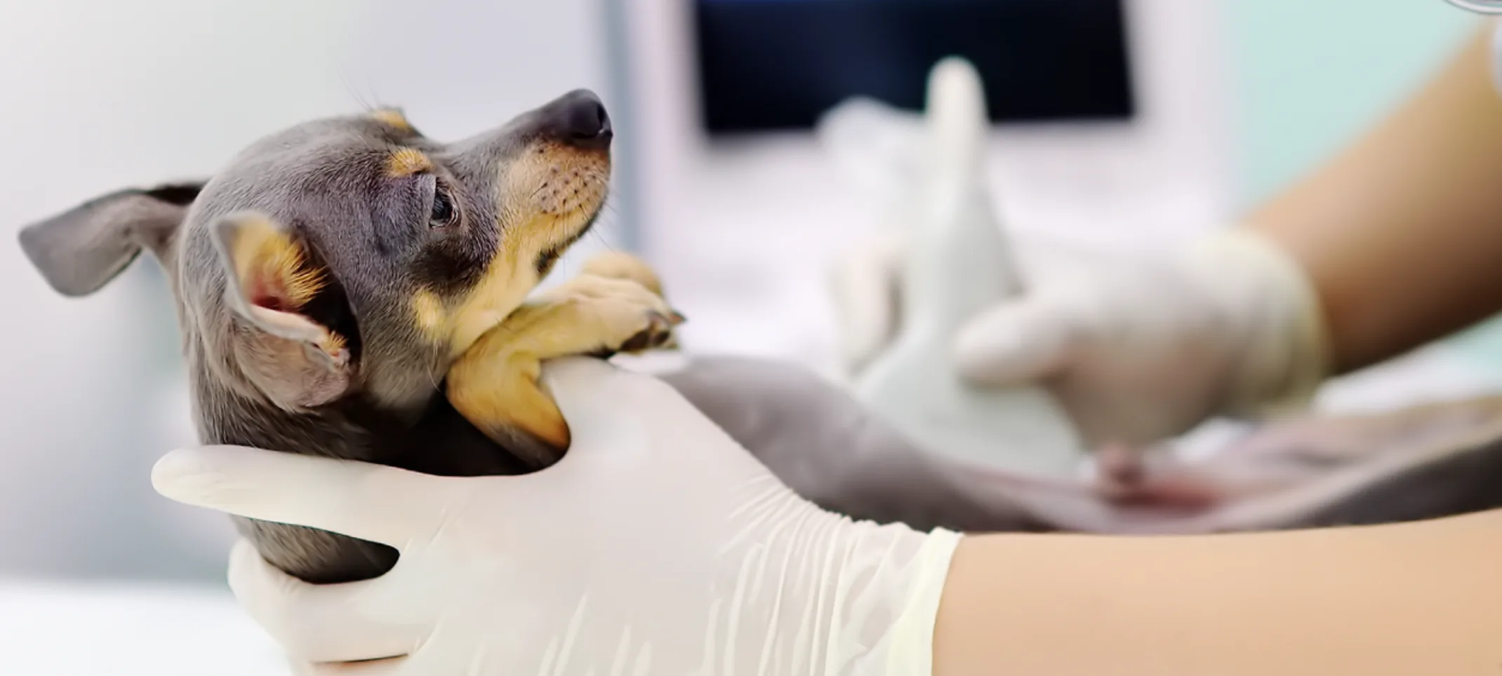
A veterinary ultrasound is an important, non-invasive diagnostic tool that helps veterinarians evaluate a variety of health conditions in pets. It is commonly used to assess heart function, detect changes in abdominal organs, and identify cysts, tumors, or other abnormalities. At NAMC, we have veterinarians who are specially trained in abdominal ultrasound, echocardiogram ultrasounds, and advanced TFAST (Thoracic Focused Assessment with Sonography for Trauma) and AFAST (Abdominal Focused Assessment with Sonography for Trauma) techniques. These specialized skills allow us to provide precise, thorough care—especially for critical cases, trauma, or emergency situations. Ultrasounds are often used in combination with x-rays, which help assess the size, shape, and position of organs, offering a more complete picture of your pet's health. Ultrasound also allows for real-time monitoring, which is particularly helpful in assessing pregnancy or tracking the development of puppies or kittens.
When Would My Pet Get an Ultrasound?
An ultrasound is typically recommended when abnormalities are detected through bloodwork, x-rays, or physical exams. It’s particularly useful for monitoring disease processes, such as kidney disease, liver issues, and heart conditions. Ultrasounds can also help assess any internal injury or trauma, evaluate organ function in detail, and assist in diagnosing conditions that require real-time imaging. These capabilities make ultrasound an essential tool for both routine health assessments and emergency care.
Advanced Diagnostic Support
At NAMC, we are committed to providing the highest quality care for your pet. We partner with Oncura, a leader in remote veterinary ultrasound consultations, connecting us with board-certified specialists in internal medicine, cardiology, and radiology. Their expert reviews of ultrasound images help guide faster, more accurate decisions, allowing us to expedite diagnosis and treatment, ensuring your pet receives the best possible care.
How Does an Ultrasound Work?
Ultrasound technology works by sending high-frequency sound waves into the body, which bounce off tissues and organs. These sound waves are reflected back and converted into images on a monitor, creating a detailed, 2D view of the internal structures. Because ultrasounds provide real-time feedback, they allow veterinarians to monitor changes immediately and make timely decisions.
The procedure is completely painless. In some cases, light sedation may be used to ensure your pet stays relaxed and comfortable during the scan. To achieve the best image quality, a small area of fur may need to be shaved so the ultrasound probe can make direct contact with the skin.
